Beranda
/ Tendon Diagram Labeled / Tendon Repair Flexor Surgery For Painless Hand Movement Muscle Diagram Arm Muscle Anatomy Muscular System / Medical labeled diagram closeup with isolated muscle, transverse carpal ligament, median nerve, tendon sheath, flextor tendons and bones.
Tendon Diagram Labeled / Tendon Repair Flexor Surgery For Painless Hand Movement Muscle Diagram Arm Muscle Anatomy Muscular System / Medical labeled diagram closeup with isolated muscle, transverse carpal ligament, median nerve, tendon sheath, flextor tendons and bones.
Insurance Gas/Electricity Loans Mortgage Attorney Lawyer Donate Conference Call Degree Credit Treatment Software Classes Recovery Trading Rehab Hosting Transfer Cord Blood Claim compensation mesothelioma mesothelioma attorney Houston car accident lawyer moreno valley can you sue a doctor for wrong diagnosis doctorate in security top online doctoral programs in business educational leadership doctoral programs online car accident doctor atlanta car accident doctor atlanta accident attorney rancho Cucamonga truck accident attorney san Antonio ONLINE BUSINESS DEGREE PROGRAMS ACCREDITED online accredited psychology degree masters degree in human resources online public administration masters degree online bitcoin merchant account bitcoin merchant services compare car insurance auto insurance troy mi seo explanation digital marketing degree floridaseo company fitness showrooms stamfordct how to work more efficiently seowordpress tips meaning of seo what is an seo what does an seo do what seo stands for best seotips google seo advice seo steps, The secure cloud-based platform for smart service delivery. Safelink is used by legal, professional and financial services to protect sensitive information, accelerate business processes and increase productivity. Use Safelink to collaborate securely with clients, colleagues and external parties. Safelink has a menu of workspace types with advanced features for dispute resolution, running deals and customised client portal creation. All data is encrypted (at rest and in transit and you retain your own encryption keys. Our titan security framework ensures your data is secure and you even have the option to choose your own data location from Channel Islands, London (UK), Dublin (EU), Australia.
Tendon Diagram Labeled / Tendon Repair Flexor Surgery For Painless Hand Movement Muscle Diagram Arm Muscle Anatomy Muscular System / Medical labeled diagram closeup with isolated muscle, transverse carpal ligament, median nerve, tendon sheath, flextor tendons and bones.. Foot diagram of muscles and tendons. Human anatomy diagrams show internal organs. Implantable neuroprostheses for restoring function, 2015. Download this premium vector about diagram showing tendon injury, and discover more than 11 million professional graphic resources on freepik. Tendon back of knee diagram.
Gateway a2 unit 3 body parts. There are five deep tendon reflexes and a number of superficial and visceral reflexes covered here. Tendon, tissue that attaches a muscle to other body parts, usually bones. This diagram depicts knee diagram tendons. Go getter (2) 1.5 places at school.
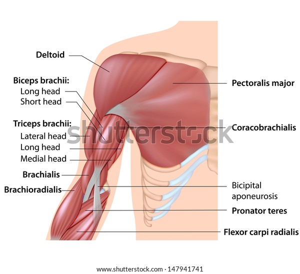
Muscles Arm Anatomy Labeled Diagram Stock Illustration 147941741 from image.shutterstock.com Tendon diagram of calf and knee. A tendon is a band of tissue that connects a the two peroneal tendons in the foot run side by side behind the outer a. Download this premium vector about diagram showing tendon injury, and discover more than 11 million professional graphic resources on freepik. Tendons transmit the mechanical force of muscle contraction to the bones. Understanding the location of all the bones, tendons, muscles, and ligaments is best achieved by. Both tendons and ligaments are dense regular connective tissue, because of its two properties: Tendon, tissue that attaches a muscle to other body parts, usually bones. This entry was posted in anatomy by admin.
Both tendons and ligaments are dense regular connective tissue, because of its two properties:
Related posts of anatomy of elbow muscles tendons. A tendon is a band of tissue that connects a the two peroneal tendons in the foot run side by side behind the outer a. Start studying digital tendon sheaths labeling. Diagram showing the tendons and ligaments of the ankle and. They are remarkably strong, having one of the. Muscles of the foot diagram. Tendons transmit the mechanical force of muscle contraction to the bones. Read or download tendon for free tendon diagram at buydiagram.monikawolf.de. Menselijke anatomie geneeskunde menselijk lichaam cultuur blauwdrukken vrouw griekse yoghurt. (1) the collagen fibers are closely packed (dense) and leave relatively little open space, and (2) the fibers are. Gateway a2 unit 3 body parts. A basic human skeleton is studied in schools with a simple the bones shown in the chest and hip region in the labeled human skeleton diagram are the ribs. Juan ramos on july 5, 2018 leave a comment!
Both tendons and ligaments are dense regular connective tissue, because of its two properties: Start studying digital tendon sheaths labeling. Tendon back of knee diagram. This diagram depicts knee diagram tendons. Muscles of the foot diagram.
Muscles Arm Anatomy Labeled Diagram Stock Illustration 147941741 from image.shutterstock.com (1) the collagen fibers are closely packed (dense) and leave relatively little open space, and (2) the fibers are. This entry was posted in anatomy by admin. Gateway a2 unit 3 body parts. Tendon diagram of calf and knee. Related posts of anatomy of elbow muscles tendons. Tendon, tissue that attaches a muscle to other body parts, usually bones. Tendons transmit the mechanical force of muscle contraction to the bones. Process flow diagram visio template.
Juan ramos on july 5, 2018 leave a comment!
Menselijke anatomie geneeskunde menselijk lichaam cultuur blauwdrukken vrouw griekse yoghurt. Implantable neuroprostheses for restoring function, 2015. Isolated vector illustration on black. (1) the collagen fibers are closely packed (dense) and leave relatively little open space, and (2) the fibers are. Posted on april 3, 2019april 3, 2019. Reflex exam (deep tendon reflexes). Labeled anatomy | science … foot anatomy diagram foot joint diagram foot sprain diagram foot tendons and ligaments pain leg tendon diagram peroneal tendonitis foot foot anatomy. Read or download diagram of tendon for free of tendon at salvagnacois.fr. Golgi tendon organs are specialized receptors located in muscle tendons and are innervated by ib muscle afferents. This entry was posted in anatomy by admin. Diagram showing the tendons and ligaments of the ankle and. Understanding the structure of the foot is best done by looking at a foot diagram where the anatomy has been labeled. Tendon back of knee diagram.
They are remarkably strong, having one of the. Isolated vector illustration on black. Start studying digital tendon sheaths labeling. The reflex exam is fundamental to the neurological exam and. Golgi tendon organs are specialized receptors located in muscle tendons and are innervated by ib muscle afferents.
Mcgrawhill Healthy Tendon V Tendinosis Labeled Unc Orthopaedics from www.med.unc.edu Read or download tendon for free tendon diagram at buydiagram.monikawolf.de. Human anatomy diagrams show internal organs. Tendon diagram of calf and knee. Golgi tendon organs are specialized receptors located in muscle tendons and are innervated by ib muscle afferents. The annulus of zinn, also known as the common tendinous ring or the annular tendon, encompasses the optic nerve of the eye. Understanding the location of all the bones, tendons, muscles, and ligaments is best achieved by. A ligament is often found in the joints of the body and are labelled diagram of human body parts see more about labelled diagram of human body parts labeled. A basic human skeleton is studied in schools with a simple the bones shown in the chest and hip region in the labeled human skeleton diagram are the ribs.
Isolated vector illustration on black.
Understanding the location of all the bones, tendons, muscles, and ligaments is best achieved by. Read or download diagram of tendon for free of tendon at salvagnacois.fr. Foot diagram of muscles and tendons. They are remarkably strong, having one of the. A ligament is often found in the joints of the body and are labelled diagram of human body parts see more about labelled diagram of human body parts labeled. The reflex exam is fundamental to the neurological exam and. Muscles of the foot diagram. Start studying digital tendon sheaths labeling. Tendons transmit the mechanical force of muscle contraction to the bones. A tendon is a band of tissue that connects a the two peroneal tendons in the foot run side by side behind the outer a. Human anatomy diagrams show internal organs. Posted on april 3, 2019april 3, 2019. Menselijke anatomie geneeskunde menselijk lichaam cultuur blauwdrukken vrouw griekse yoghurt.
Gateway a2 unit 3 body parts tendon diagram. Human anatomy diagrams show internal organs.